Main Second Level Navigation
- Bachelor of Science in Medical Radiation Sciences
- Medical Physics Residency
- Radiation Oncology Residency
- Radiation Oncology Fellowship
- Visiting and Elective Residents and Fellows
- Clinical and Experimental Radiobiology Course
- MR-integrated Radiation Therapy Training Program
- Learner Mistreatment
- Electives and Observerships
- MSc and PhD
- Student Life and Resources
- STARS21
- Teaching Evaluations
Breadcrumbs
- Home
- Education & Continuing Education
- Bachelor of Science in Medical Radiation Sciences
- Radiological Technology
Radiological Technology
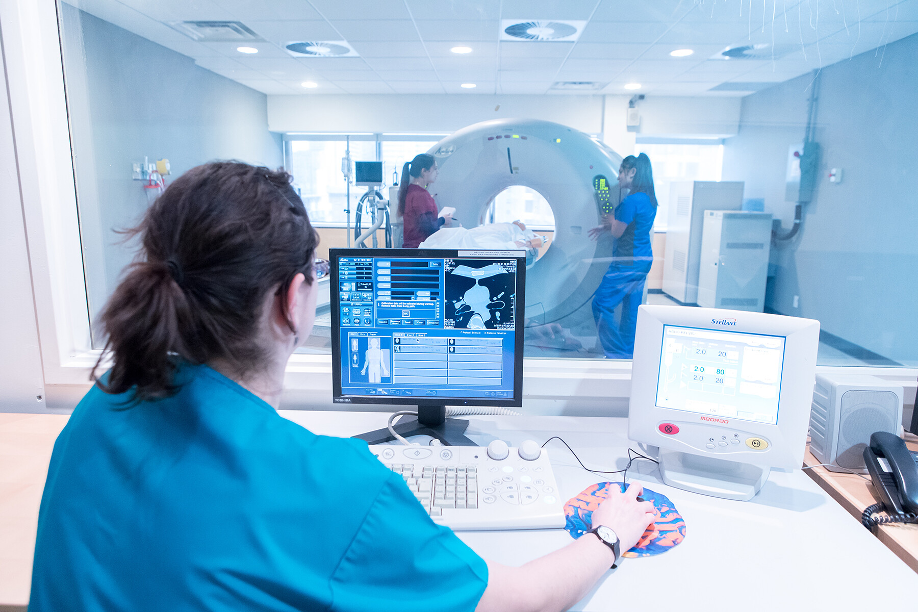
Radiological technology is the production of medical images, commonly called X-rays, of internal organs and structures. They are produced by passing a small, highly controlled amount of radiation through the human body, and capturing the resulting image on an image recording device.
To enhance certain organs and structures that otherwise are not visualized on medical X-ray image, contrast media may be used.
The field of radiological technology includes many different subspecialties. Examples include the following:
- General Radiology is used to detect bone fractures and pathological processes, locate foreign objects in the body, and demonstrate the relationship between bone and soft tissue.
- Fluoroscopy produces real-time X-ray images. Fluoroscopy is used in conjunction with contrast media to enable visualization of internal structures such as the gastrointestinal tract, blood vessels and various organs during diagnostic and therapeutic procedures. Fluoroscopy is also performed in the operating room during surgery providing the surgeon visual guidance for various surgical procedures.
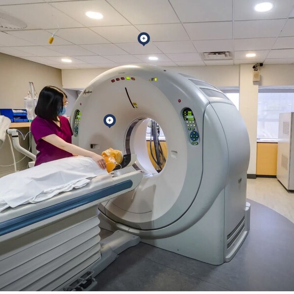
- Computerized Tomography (CT) produces cross-sectional and 3-dimensional images of various structures in the body
- Angiography uses contrast agents to examine the heart and blood vessels
- Mammography produces radiographic images of the breast to detect cancer in its earliest stages
- Electronic image management (PACS)
What does a Radiological Technologist do?
Radiological Technologists are vital members of the interprofessional health care team devoted to patient care. Technologists have the technical expertise to operate sophisticated instruments, and also have the humanistic skills necessary to communicate with patients, problem-solve, and work well with other members of the health care team. Radiological Technologists will:
- Play an integral role in the detection of injury and disease; they are the medical personnel who perform diagnostic imaging examinations, including mammography and computerized tomography.
- They are responsible for accurately positioning patients and ensuring that a quality diagnostic image is produced.
- Use cutting-edge medical imaging technology and advanced computer systems to perform complex anatomical scans, many in real-time, to produce and enhance radiographic images.
- Interventional radiography is use for the detection, diagnosis and treatment of injury and disease.
Is Radiological Technology a good choice for you?
Radiological Technologists enjoy a variety of hospital settings and roles including the fast-paced emergency and operating room environments, as well as private clinics, research institutes and public health institutions. As a Radiological Technologist, you are an integral member of a patient’s diagnostic pathway, where you may specialize in a particular area of diagnostic imaging such as:
- Mammography (breast imaging)
- Computed tomography (CT)
- Diagnostic visceral and peripheral angiography with interventional radiology
- Electronic image management (PACS)
- Neuroradiology or trauma radiography
Your future career opportunities could include computed tomography (CT), management, education and sales/marketing, magnetic resonance imaging (MRI), research activities, medical equipment sales, government policy, as well as pursuing higher education opportunities at a master’s level, and working abroad.
Graduates of the Radiological Technology stream of the MRS program will earn a BSc in Medical Radiation Sciences from the University of Toronto and an Advanced Diploma in Health Sciences (Radiological Technology) from The Michener Institute. MRS graduates are eligible to write the national certification exam offered by the Canadian Association of Medical Radiation Technologists (CAMRT) and may pursue advanced studies at U of T or Michener, such as:
- Magnetic Resonance Imaging (MRI)
- Master of Applied Science (Institute of Medical Sciences)
- Master of Health Administration
MRS Clinical Placements - Radiological Technology
Radiological Technology students gain experience through non-paid clinical placements at one or more sites primarily within the Greater Toronto Area including: Ajax/Pickering, Barrie, Markham, Oshawa, Richmond Hill, and Toronto*. Radiological Technology students graduate with 42 weeks of clinical experience:
- 8 weeks at the end of Year 1
- 4 weeks at the end of Year 2
- 30 weeks in Year 3
* Please note that placement at your first-choice site is not guaranteed and that clinical site locations are subject to change.
MRS Course List*- Radiological Technology
*Subject to Change
YEAR 1 FALL – SEMESTER 1
MRS159H1 / ANAT110 – Anatomy for Medical Radiation Sciences
This course is an introductory online course designed to serve as the foundation in Human Anatomy for students in the Medical Radiation Sciences program. The course will introduce learners to the components of the human body, relationships of the surface anatomy and the body's internal components and discuss the basic function of these components. The course will encompass a regional approach to study the human body with correlation to its clinical application. Anatomy for MRS will also serve to prepare learners for ANRD121/ MRS164H1 (Relational Anatomy).
MRS102H1 / ANRA310 – Human Osteology
The course will focus on the study of human osteology. Labs are structured for small group learning where learners have the opportunity to examine anatomic models in gaining a comprehensive understanding of skeletal anatomy which is vital for the practice of Radiological Technology. Learners will be guided by the instructor in identifying the anatomic features listed on the learning activity for each bone of the appendicular and axial skeleton.
Learners will also apply their learning when viewing radiographic images and indetifying the bony features that are visible on various radiographic projections. Palpable bony landmarks will also be reinforced in discussing accuracy of positioning.
Selected ligaments of the common joints that are imaged by X-Ray will also be studied using anatomic models and drawings.
MRS281H1 / IGRD110 – Comparative Medical Imaging
This course is designed to introduce the learner to the complexities of diagnostic and therapeutic imaging in the healthcare setting. Learners will develop an understanding of what it means to be a medical radiation technologist, a healthcare professional, and interprofessional collaboration. As well, learners will be exposed through a series of seminars delivered by healthcare professionals from the practice setting, to a variety of imaging modalities used in the diagnosis and treatment of patients. Modalities such as X-ray, Computed Tomography (CT), Magnetic Resonance Imaging (MRI), Ultrasound (US), Positron Emissions Tomography (PET), and Image Guidance for Radiation Therapy will be explored. Additional topics relating to the imaging modalities will be covered including informatics, Artificial Intelligence/Machine Learning and Interventional Radiology.
MRS227H1 / PMRS111 – Patient Care in Medical Radiation Sciences I
Examining a variety of fundamental patient care topics using a patient-centred lens, learners will be introduced to the professional, ethical, and legal standards which will be reinforced throughout the Medical Radiation Sciences (MRS) curriculum. Patient care skills will be enhanced through the study of communication, medical terminology, infection control, clinical assessment techniques and relevant pharmacology, including contrast media. This course will also provide the opportunity to develop communication and care-delivery strategies for diverse patient populations. Reflective practice will be introduced and developed throughout the course as learners work towards becoming reflective practitioners.
MRS114H1 / INRA110 – Diagnostic Imaging Instrumentation I
The purpose of this course is to introduce learners to the principles of medical image recording and equipment. In this course, learners will gain introductory knowledge on how radiological images are produced, manipulated, and critiqued in terms of diagnostic quality. Learners will examine how information flows from the patient to the observer in four distinct stages.
1. Formation of the invisible/latent image, in both analogue and digital format
2. Conversion of the invisable/latent image into a visible radiographic image in both analogue and digital format.
3. Examination of X-ray quality and quantity and their effect on the radiographic image.
4. Analysis and perception of the radiographic image.
MRS115H1 / PHRA110 – Radiographic Physics and Radiobiology
The Radiographic Physics portion of this course describes the properties and components of the electromagnetic spectrum. Medical X-ray production and equipment will be discussed, along with the factors that determine both the quality and quantity of the X-ray beam. The course also examines the interactive processes between matter and the X-Rays. As well, the principles relating to detection and quantification of ionizing radiation are examined. Finally, the fundamental principles of radiation safety in the practice of radiological technology will be applied.
The Radiobiology portion of this course will be delivered on line and jointly with the students of the Nuclear Medicine and Molecular Imaging Technology program. This course examines the biological response to radiation in depth, ranging from the cellular level to whole body response.
YEAR 1 WINTER – SEMESTER 2
MRS164H1 / ANRD121 – Relational Anatomy
For the purposes of diagnosis and optimal treatment of abnormal patient conditions, high quality, detailed images are required in order to obtain information on the anatomical state of an organ or structure within the body. The ability to recognize anatomical structures as demonstrated on radiographic images and in sagittal, coronal and axial sectional planes generated by computed tomography and magnetic resonance imaging is essential in the current hospital imaging environment.
This Relational Anatomy course requires students to apply their knowledge of gross anatomy to explore the cross-sectional and relational anatomy of the head, central nervous system, neck, spine, thorax, abdomen, pelvis (male/female) and the upper and lower extremities. An emphasis is placed upon the student’s ability to identify and justify the relative positions of the organs, the vascular system, the lymphatic system as well as muscular and skeletal structures in each of the aforementioned anatomic regions. This course offers weekly interactive lectures and labs which provide the student with a stimulating environment in which they may actively apply the knowledge learned through the use of a variety of media including anatomical models, preserved human specimens, cadaver cross section images as well as a multitude of hard copy and computer based medical images.
MRS228H1 / PMRS121 – Patient Care in Medical Radiation Sciences II
In this course, learners will build upon the knowledge acquired in PMRS111 as they apply the concepts of professional practice, patient management and health and safety, in both the didactic and laboratory environments. Learners will employ principles of infection control, patient assessment, patient transfer, contrast media administration and emergency response procedures. Learners will also be introduced to various venipuncture techniques. These practical skillsets will be developed as learners simultaneously integrate the concepts of patient-centred communication and professionalism introduced in PMRS111.
MRS162H1 / PSRD120 – Physiology
This course is an introductory ONLINE course designed to serve as the foundation in Human Physiology for students in the Medical Radiation Sciences program. It is intended for students who have an interest in or a need for a basic course in Human Physiology. The course is 0.5 credit course and will introduce students to the function of the organ systems that comprise the human body. The course will follow a systematic structure covering all of the principal functional systems within the body, such as the cardiovascular and respiratory systems. As such students are expected to be familiar with the anatomical structure of these systems. Clinical examples will be used to illustrate key principles and material where possible.
MRS116H1 / INRA120 – Diagnostic Imaging Instrumentation II
This course is a continuation of the concepts and practice learned in Diagnostic Instrumentation I. Building upon previous knowledge, the learner will continue to gain insight into the principles of medical image recording, and will further explore the modalities of CR and DR, in greater depth. The link between Digital Imaging and Picture Archiving and Communication System (PACS) will be emphasized, and the relationship between Hospital Information System (HIS), Radiology Information System (RIS) and PACS will be explored.
In addition to classroom based sessions which emphasize the theory behind obtaining optimum diagnostic quality medical images, students will work through problem solving activities in the laboratory setting to reinforce theory. Students should be able to discuss how the results of each laboratory activity cn be directly applied to the clinical setting.
MRS233H1 / RARA311 – Radiographic Procedures I
Radiographic Procedures I focuses on aiding learners to achieve the skills necessary to safely and correctly complete radiographic studies of the appendicular skeleton, chest and abdomen. The fundamental principles of radiographic image production, the ability to analyze radiographic images for correct positioning and technical factor selection, and the parameters that define optimal diagnostic quality of a radiographic image will be introduced in this course, in addition, the importance of radiation protection principles and practices will be reinforced.
YEAR 1 SUMMER – SEMESTER 3
MRS118H1 / CLRA130 – Introduction to Clinical Radiography
In this course the learner will gain an understanding of the role and responsibility of the MRT in the clinical environment. While in the clinical setting, the learner will have an opportunity to practice general radiography in their assigned clinical site. The learner will gain an understanding of the patient experience by applying the patient care, communication, and methodology skills learned in the didactic environment to the practice of Radiological Technology. They will familiarize themselves with the mechanical functioning of the imaging equipment and experience the team dynamics of health care provision. As the learner becomes increasingly familiar with the role of the MRT he/she should begin to take on a more participatory role. Further to the time spent in general radiography, the learner, where possible, may have the opportunity to spend a minimum of one day (maximum of three days) in one or more of the following areas: computed tomography (CT), portable/operating room (OR) radiography, gastric imaging, BMD, and interventional radiography. Learners will also have the opportunity to visit the Mammography department to examine the imaging equipment in preparation for INRA251/MRS205H1 – Diagnostic Imaging Instrumentation course.
Selective I
The specialized electives (“selectives”) are courses developed with the purpose of providing graduates of the Medical Radiation Sciences (MRS) Program with knowledge and expertise in specialized fields of practice. Selectives are designed to give students some freedom in constructing a curriculum that responds to their own particular interests related to their chosen profession and/or academic endeavors after graduating from the program.
Examples of selective courses that are available to MRS students are: MRI, Informatics, Mammography, Patient Education, Supportive and Palliative Care, and many more.
YEAR 2 FALL – SEMESTER 4
MRS266H1 / RMIP240 – Introduction to Research Methods
This course provides an introduction to research methods and designs relevant to practitioners of the medical radiation sciences. This course will focus on an introduction to various research designs including experimental and non-experimental, and quantitative and qualitative research methods. In addition, the course will focus on providing a practical understanding of several basic statistical tools used in medical and health research.
MRS265H1 / CTRD240 – Integrated C.T. Imaging Theory and Practice I
This course emphasizes the delivery of safe, efficient CT imaging in a manner using best practices to care for patients. The course familiarizes the students with the working components of a CT scanner, the theory behind the acquisition of CT images and the manipulation of the various technical parameters required to complete a head, chest, abdomen, pelvis and skull CT scan. Using a CT phantom, the learners will participate in simulated clinical applications of computed tomography with a focus on dose management and optimization.
MRS205H1 / INRA251 – Diagnostic Imaging Instrumentation III
This course provides an introduction to all aspects of Quality Assurance and Quality Control of Radiographic Imaging Equipment. Beginning with a review of the factors affecting image quality, the topics of Quality Management, Legislation and Continuous Quality Improvement will be discussed. The focus of this course will be quality control of image acquisition and display equipment. After acquiring fundamental theory in lectures, and through self-directed course preparation, students will have the opportunity to perform quality control testing on diagnostic X-ray imaging equipment in the laboratory setting.
MRS253H1 / RARA241 – Radiographic Procedures II
Radiographic Procedures II focuses on aiding learners to achieve the skills necessary for safely and correctly completing radiographic studies of the axial skeleton and thorax. The importance of radiation protection principles and practices will be reinforced. The ability to correctly position the patient, selection of technical factors and parameters that define optimal diagnostic quality of a radiographic images will continue to be developed. This course is a combination of both theory (classroom based) and practice (laboratory).
MRS254H1 / RARA242 – Radiographic Image Analysis
This course focuses on aiding learners to develop image analysis skills while correlating accurate technical and positioning procedures learned in Radiographic Procedures II. Students will further expand their image assessment skills of the chest and abdomen. Students will be introduced to image assessment of the axial skeleton and bony thorax.
Students will assess images for correct positioning, technical factor selection and parameters that define optimal diagnostic quality of a radiographic image. Students will determine the acceptability of images or exams with respect to patient clinical history and recommend any corrective measures. The importance of radiation protection principles and practices will be reinforced. This course is a combination of both theory and practical application.
MRS260H1Y / EMRS240 – Experiential Learning in IPEC
"lnterprofessional education (IPE) occurs when students from two or more professions learn about, from and with each other to enable effective collaboration and improve health outcomes" (WHO, 2010). At the academic level, the goal of IPE is to prepare health professional students with the knowledge, skills and attitudes necessary for collaborative interprofessional practice (http://ipe.utoronto.ca/).
Experiential Learning in lnterprofessional Education and Collaboration is a longitudinal, two semesters, course consisting of online asynchronous and synchronous learning activities. Online asynchronous self-directed activities will explore and examine key concepts and skills surrounding IPE, reflection, and communication.
Synchronous experiential learning activities will expose students to other health profession students in various contexts to enable them to better understand health profession roles, appreciate team dynamics in healthcare, and learn with and from each other. Through the various learning opportunities, MRS students will gain valuable collaborate and communication skills to help prepare them to be collaborative practice-ready healthcare professionals. This course is interactive and requires full participation. Students will be required to reflect on course content and activities and prepare a longitudinal portfolio that reflects the values and competencies of interprofessional education and collaboration.
YEAR 2 WINTER – SEMESTER 5
MRS269H1 / HBRD241 – Clinical Behavioural Sciences
This course provides an overview of the ethical, societal and personal factors that influence and impact health care services. The role of the health care provider and the dynamics of the provider/patient relationship will be examined from a critical perspective. Through an interprofessional education session students will explore alternate models of healthcare provision. This course will combine theory and practical application, allowing the student to reflect on his or her own values and beliefs through case studies, reading and discussion.
MRS206H1 / CTRD250 – Integrated C.T. Imaging Theory and Practice II
Computed Tomography (CT) is one of the essential competencies required for an entry-level technologist for Radiological Technology in Canada. This course is designed to ensure that students recognize the appearance of common pathologic conditions and anomalies seen on CT scans of the head, neck, chest, abdomen, pelvis, extremities and spine. This course will explore the significance of CT procedures and protocols used to visualize common pathologic conditions and anomalies on CT images. In a simulated setting in the CT suite the learner will manipulate scan protocols and the acquired image data from a prescribed study in preparation for diagnostic reporting.
MRS256H1 / RARA250 – Radiographic Procedures III
This course is a study in advanced radiographic and fluoroscopic imaging procedures of the body systems. In the previous methodology courses RARA311 and RARA240, the technologist (as the primary practitioner) focuses on their positioning skills in performing general radiographic imaging examinations. In the performance of advanced diagnostic and interventional imaging examinations covered in this course, the technologist’s role is that of an essential member of the Medical Imaging health care team and is responsible for the operation of specialized equipment, room preparation for the procedure, patient monitoring and assisting the radiologist during the procedure. Methodology III will also explore the importance and application of contrast media in the visualization of the gastrointestinal, genitourinary, reproductive, musculoskeletal, central nervous and cardiovascular system. This course will enhance the fundamental knowledge and skills acquired in Radiographic Methodology I and II pertaining to positioning, patient care, image assessment, and radiation protection. Anatomical variants and pathology pertaining to the body system covered in this course will be discussed. This course is a combination of both theory (classroom based) and laboratory sessions. All Lectures and Labs will be held online and conducted in a virtual format.
MRS119H1 / PGRA240 – Medical Imaging Pathology
This course provides the student with the basic knowledge to understand common pathological conditions and anomalies and to recognize them on radiographic images. Patient signs and symptoms as well as clinical presentations will be discussed during the course. Technical factors required to best visualize these pathologies and anomalies will be discussed.
MRS212H1 / PCRA251 – Patient Care for Medical Imaging
This course focuses on patient care specific to areas of practice in Radiological Technology. Patient management in the areas of Trauma, ICU, OR and special patient considerations will be investigated.
MRS260H1Y / EMRS240 – Experiential Learning in IPEC (Continued)
"lnterprofessional education (IPE) occurs when students from two or more professions learn about, from and with each other to enable effective collaboration and improve health outcomes" (WHO, 2010). At the academic level, the goal of IPE is to prepare health professional students with the knowledge, skills and attitudes necessary for collaborative interprofessional practice (http://ipe.utoronto.ca/).
Experiential Learning in lnterprofessional Education and Collaboration is a longitudinal, two semesters, course consisting of online asynchronous and synchronous learning activities. Online asynchronous self-directed activities will explore and examine key concepts and skills surrounding IPE, reflection, and communication.
Synchronous experiential learning activities will expose students to other health profession students in various contexts to enable them to better understand health profession roles, appreciate team dynamics in healthcare, and learn with and from each other. Through the various learning opportunities, MRS students will gain valuable collaborate and communication skills to help prepare them to be collaborative practice-ready healthcare professionals. This course is interactive and requires full participation. Students will be required to reflect on course content and activities and prepare a longitudinal portfolio that reflects the values and competencies of interprofessional education and collaboration.
YEAR 2 SUMMER – SEMESTER 6
MRS209H1 / CLRA261 – Simulated Clinical Practice: Radiological Technology
This is a 12-week clinical simulation course designed to prepare the learner to enter into the clinical education environment of a medical imaging department. The learner will have the opportunity to integrate technical and professional competencies required for the practice of radiography. Each learner will be engaged for 12 hours per week in the following learning activities:
• Selected Topics in Radiological Technology
• Image Acquisition and Assessment
• Case Study of Clinical Scenarios
• General Radiography Practice
• Patient Care and Related Radiographic Imaging Procedures
MRS231H1 / TCRA266 – Transitional to Clinical Radiography
This transition course is designed to provide students with opportunity to further enhance their knowledge, skills and judgement from the didactic and clinical simulation (CLRA261/MRS209H1) environments. As they transition into clinical radiological technology practice and the year 3 clinical courses, the learners’ experience will concentrate primarily on the practice of general radiography (upper and lower extremities, respiratory/chest, and abdominal imaging).
MRS232H1 / HIRT130 – Health Improvement Initiatives
Health Improvement Initiatives is a 12-week summer semester course that will focus on the intersection of three principle domains of healthcare system, leadership, quality. The course will explore the organization and structure of the Canadian healthcare system to better understand and appreciate the roles and accountabilities of those who use and those who function within it. It will also examine healthcare leadership at all levels and the practitioner needed for today’s dynamic healthcare environment. Lastly, the course will focus on understanding quality improvement initiatives aimed to make healthcare more effective, efficient, and safe.
Selective II
The specialized electives (“selectives”) are courses developed with the purpose of providing graduates of the Medical Radiation Sciences (MRS) Program with knowledge and expertise in specialized fields of practice. Selectives are designed to give students some freedom in constructing a curriculum that responds to their own particular interests related to their chosen profession and/or academic endeavors after graduating from the program.
Examples of selective courses that are available to MRS students are: MRI, Informatics, Mammography, Patient Education, Supportive and Palliative Care, and many more.
YEAR 3 FALL – SEMESTER 7
Clinical Radiography II (CLRA371/MRS210H1)
This fifteen week clinical course provides the learner the opportunity to put into practice their knowledge of radiographic and fluoroscopic imaging procedures of the body systems. The learner will have the opportunity to further acquire and integrate knowledge, skills, attitudes and judgement required for the practice of clinical radiography. Clinical rotations are required of the learner in order to achieve competency in the practice of clinical radiography.
A series of supervised clinical courses in affiliated clinical sites form the basis of clinical education for all three disciplines of the Medical Radiation Sciences (MRS) program. The clinical courses provide the student with the opportunity to develop and demonstrate the expected professional skills and behaviours, and to integrate the requisite knowledge to achieve competence in all aspects of the chosen discipline, including those outlined in published documents of the certifying and licensing associations. Staff professionals and Clinical Coordinators work to guide and assist the student in meeting these goals.
RMRD370 / MRS398Y1 – Research Methods
This two-semester course is the practical component of the Introduction to Research Methods course (MRS265H1/RMIP240). It allows for the completion of a clinically based research project under supervision. The research question and subsequent project may be pre-determined by the clinical research supervisor as it will most likely pertain to the supervisors' research area of interest and clinical expertise. Students will be matched with a research supervisor at their clinical host site to facilitate the development of a clinically relevant and feasible research study.
Delivery and support of this course is principally on-line and at a distance. However, students may be required to present assignments in-person or via live-electronic media in the winter semester of the course. The opportunity to meet in-person or on-line will assist in establishing a collegial research network and community amongst the learners. Completion of the research project will culminate in the submission of a written research thesis. Students will be encouraged to complete work of publishable quality.
This course is recommended for students who are interested in pursuing graduate studies and/or an academic career in Medical Radiation Sciences
- OR -
MRS397H1 / RPRD370 – Research in Practice
Research in Practice is a one semester online course designed to provide an opportunity for learners to critically examine the role that research plays in informing and advancing practice. Through an inquiring examination of clinical practice, the learner will gain an appreciation for evidence-based practice and research. The course consists of independent self-directed assignments in which the learner will review and critically appraise a procedure/guideline from practice (imaging, therapeutic, patient, safety), generate questions and rationale for further investigation, and conduct a search of current literature that leads to a written review of evidence-based practice.
YEAR 3 WINTER – SEMESTER 8
MRS211H1 / CLRA380 – Clinical Radiography III
This is the final clinical course. It provides the student with the final opportunity to develop the skills and behaviours practiced in MRS210H1/CLRA371. The student will be provided with a wide variety of experiences in the clinical setting, in all professional behaviour situations, they are expected to meet the expectations of an entry level practitioner. The student in this course is expected to demonstrate competence in all the required skills as outlined in the CAMRT Competency Profile and Summary of Clinical Competence. Staff professionals and Clinical Coordinators work with the student to facilitate the achievement of these goals as they approach the end of their clinical experience. Supervision of the student may be by direct or indirect observation, at the discretion of the presiding clinical Coordinator or clinical staff.
RMRD370 / MRS398Y1 – Research Methods
This two-semester course is the practical component of the Introduction to Research Methods course (MRS265H1/RMIP240). It allows for the completion of a clinically based research project under supervision. The research question and subsequent project may be pre-determined by the clinical research supervisor as it will most likely pertain to the supervisors' research area of interest and clinical expertise. Students will be matched with a research supervisor at their clinical host site to facilitate the development of a clinically relevant and feasible research study.
Delivery and support of this course is principally on-line and at a distance. However, students may be required to present assignments in-person or via live-electronic media in the winter semester of the course. The opportunity to meet in-person or on-line will assist in establishing a collegial research network and community amongst the learners. Completion of the research project will culminate in the submission of a written research thesis. Students will be encouraged to complete work of publishable quality.
This course is recommended for students who are interested in pursuing graduate studies and/or an academic career in Medical Radiation Sciences
– OR –
Selective III
The specialized electives (“selectives”) are courses developed with the purpose of providing graduates of the Medical Radiation Sciences (MRS) Program with knowledge and expertise in specialized fields of practice. Selectives are designed to give students some freedom in constructing a curriculum that responds to their own particular interests related to their chosen profession and/or academic endeavors after graduating from the program.
Examples of selective courses that are available to MRS students are: MRI, Informatics, Mammography, Patient Education, Supportive and Palliative Care, and many more.

